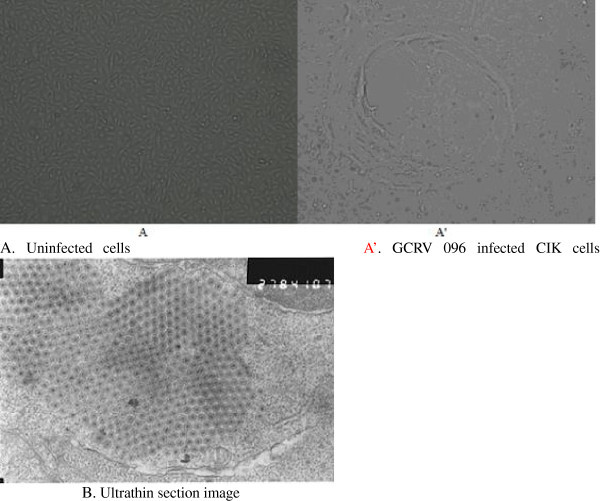
Figure 1.

CPE in the CIK cells 3 d after GCRV 096 isolate inoculation (A, A’ 100×) and Crystalline array of viral particles (B 50,000×). Notes: A. The control CIK cells without GCRV096 inoculation. A’. CPE in the CIK cells 3 d after GCRV 096 isolate inoculation. B: Crystalline array of viral particles.
