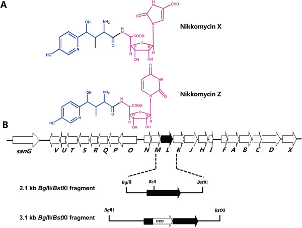
Figure 1.
Chemical structure (A) and organization of the gene cluster for nikkomycin biosynthesis (B). The peptidyl moiety (HPHT) and nucleoside moiety of nikkomycin were indicated by blue color and red color, respectively. The solid arrow shows sanL and its orientation. The 2.1 kb BglII/BstXI fragment contains sanL and its flanking sequences. The 3.1 kb BglII/BstXI fragment was used for disruption of sanL. The kanamycin resistance gene (neo) was inserted into BclI site of sanL.

