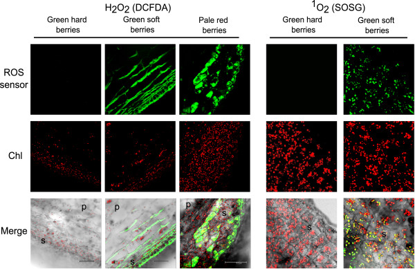
Figure 2.

Confocal images of Pinot Noir berries (100-μm sections) sampled at the green hard, green soft and pale red stages, stained for H2O2and1O2. The sections were incubated with either 30 μM DCFDA or 30 μM SOSG (ROS sensors). Chlorophyll fluorescence has been recorded (Chl) to localize chloroplasts inside the cells. Merge is the computed overlay of the two fluorescence images and the bright field. Reference bars are 75 μm for H2O2 imaging and 25 μm for 1O2. Skin and pulp are indicated in the merge pictures with a “s” and “p”, respectively. For 1O2 imaging, only skin is visualized, at a higher magnification.
