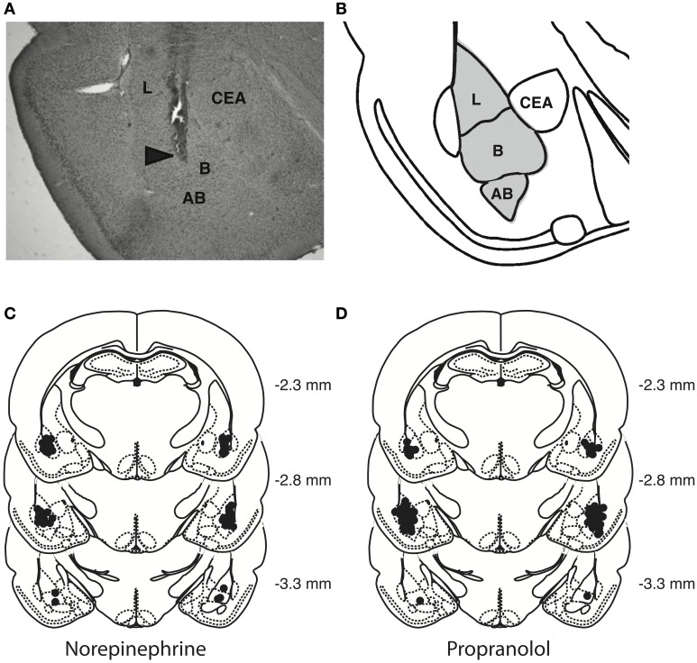
Figure 2.
Histological analyses. (A) Representative photomicrograph illustrating placement of a cannula and needle tip in the BLA. Arrow points to needle tip. (B) The gray area in the diagram represents the different nuclei of the BLA: the lateral nucleus (L), basal nucleus (B), and accessory basal nucleus (AB). CEA, central nucleus of the amygdala. (C,D) Location of infusion needle tips of all rats included in the final analyses.

