Figure 4.
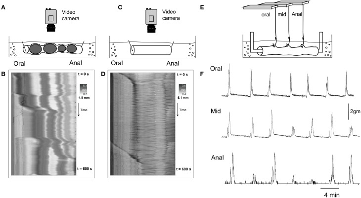
(B,D,F) are three separate recordings, from the same segment of whole mouse colon, using different recording methodologies. (A) Shows a diagrammatic representation of the preparation from which video recordings were made from a whole colon that contained multiple fecal pellets. (B) Shows the D-map from the same preparation, with CMMCs present that propel a number of pellets aborally. (C) Shows the preparation from which video recordings were made from the same segment of colon, but devoid of all fecal pellets. (D) Shows a D-map from the same preparation of colon as in (B) but the colon is now devoid of all fecal pellets. CMMCs are considerably less frequent. (E) Shows a diagram of the preparation but now when isometric force transducers are attached to the circular muscle at three sites along the colon, with 1 gm resting tension imposed. (F) Shows that under these recording conditions, CMMCs are now regularly recorded which propagated from oral, to mid, to distal colon.

