Figure 1.
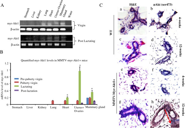
Expression of activated Akt1 in transgenic mice at different development stages. (A) Total RNA was extracted from the indicated organs of virgin (9 week old) and post-lactating (16 week old) transgenic mice. RT-PCR was performed with primers for myr-Akt1 and β-actin. Three mice per genotype, per age group were used for analysis. Myr-Akt1 was detected in the lungs, heart, uterus, and mammary glands of the virgin and post-lactating mice. (B) The mRNA levels of myr-Akt1 were quantified in different organs of the transgenic mice at different developmental states. Myr-Akt1 levels were adjusted for the levels of β-actin. Data are presented as bar graphs. The bars indicate mean levels and SD from three mice and “*” indicates p < 0.05 compared to the virgin mice. (C) H&E staining (a and b), and immunohistochemical detection of phospho-Akt1 (ser743) (c and d) in the mammary glands of wild-type mice (WT) and transgenic mice (MMTVmyr-Akt1).

