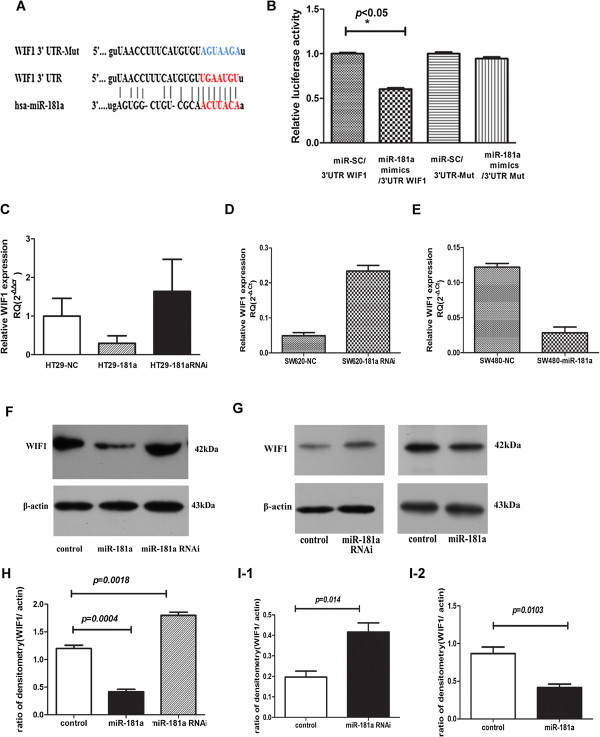
Figure 4.
WIF-1 is target of miR-181a. (A) Schematic illustration of the predicted miR-181a-binding sites in WIF-1 3′-UTR; (B) Luciferase reporter assay demonstrates that miR-181a inhibited the wild-type, but not the mutant, 3′-UTRs of WIF-1 reporter activities compared with the vector alone control. The data represent the mean ± SD of three independent experiments with quadruplicates of sample. Student’ s t-test, * p < 0.05 versus control (wild-type 3 -UTR reporter vector + miR scramble) or mutant 3-UTR reporter group (mutant 3 -UTR reporter + miR-181a mimics/miR scramble); (C) The expression of endogenous WIF-1 was inhibited in the pool of lenti-pri-181a-infected HT29 cells and enhanced in lenti-pri-181a-RNAi-infected HT29 cells, compared with the control, at mRNA level as detected by qRT-PCR; bar, mRNA expression normalized to GAPDH mRNA; (D, E) WIF-1 mRNA levels were substantially enhanced in lenti-pri-181a-RNAi-infected SW620 (D) and suppressed in SW480 overexpressing miR-181a cells(E), compared with the controls; (F, G) Western blot results show that the proteins of WIF-1 were down-regulated following lenti–pri–181a infection and up-regulated following lenti-pri-181a-RNAi infection (F, HT29 cell; G, SW620 and SW480cells). β-Actin served as an internal loading control. (H, I) The statistical analysis results of ratio of WIF1 compared to β-actin (H, HT29 cell; I-1, SW620; I-2, SW480 cells). Data represent the mean ± SD of three independent experiments.

