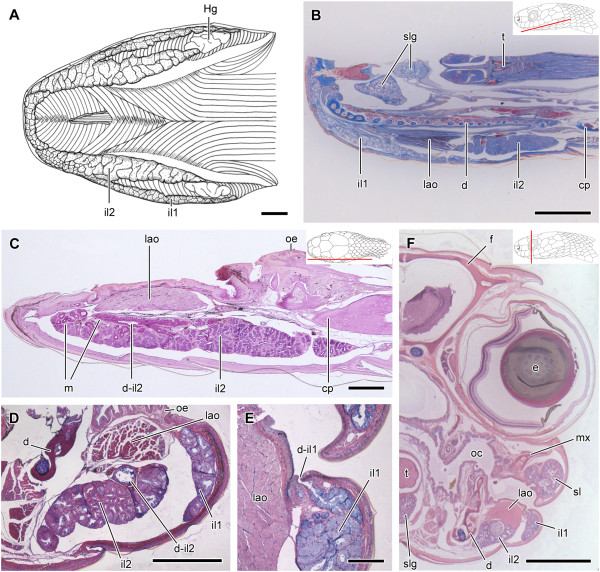
Figure 8.
Histological sections of the head of Sibynomorphus mikani and S. neuwiedi. Ventral view of the skinned head of Sibynomorphus mikanii evidencing the size and location of both il1 and il2; Paraffin section, Mallory trichrome staining (A). Sagittal section of the head of Sibynomorphus mikanii showing the position of the two portions of the infralabial gland (il1 and il2); Paraffin section, Mallory trichrome staining (B). PAS histochemical reaction in a longitudinal section of the mandibular region of Sibynomorphus mikanii (MZUSP 17886) revealing the more developed portion of infralabial gland (il2) with the duct (d-il2) running towards the anterior region. Although the whole gland reacts to PAS, mucous cells (m) in the anterior region and the duct (d-il2) are much more positive; Paraffin section (C). Transverse section of the mandibular region of Sibynomorphus mikanii (MZUSP 17882) showing the infralabial gland divided in il1 and il2, and evidencing the duct of il2 (d-il2); Paraffin section, Hematoxylin-eosin staining (D). Transverse section of the head of Sibynomorphus neuwiedi (MZUSP 17225) evidencing a duct in il1; Paraffin section, Hematoxylin-eosin staining (E). Transverse section of the head of Sibynomorphus neuwiedi showing il1 and il2 separated by the bundle of the muscle levator anguli oris; Paraffin section, Hematoxylin-eosin staining (F). Abbreviations: cp, compound bone; d, dentary bone; d-il1, ducts of the lateral, mucous infralabial gland; d-il2, duct of the ventrolateral, seromucous infralabial gland; e, eye; f, frontal; Hg, Harderian gland; il1, lateral, mucous infralabial gland; il2, ventrolateral, seromucous infralabial gland; lao, muscle levator anguli oris; m, mucous cells; mx, maxillary; oc, oral cavity; oe, oral epithelium; sl, supralabial gland; slg, sublingual gland; t, tongue. Scale bar in pictures A-D and F = 1 mm and E = 50 μm. Panels at the upper right corner denote position of the section in B, C, F.

