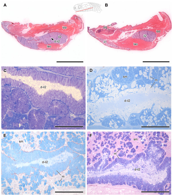
Figure 9.
Histological sections of the infralabial glands of Dipsas neivai. Two distinct horizontal planes from serial histological sections of the head of Dipsas neivai (MZUSP 14665) showing part of the larger infralabial gland (il2) with the duct (d-il2) in the central area (A), and part of the smaller and thinner mucous infralabial gland (il1), extending along the margin of the lip (B), and their relationship with muscles levator anguli oris (lao) and intermandibularis posterior pars posterior (ipp). While the larger seromucous infralabial gland (il2) is embraced by both muscles, the thinner gland (il1) is connected only with the muscle levator anguli oris; Paraffin sections, Hematoxylin-eosin staining (A-B). Longitudinal historesin sections of il2 focusing the duct (d-il2) and the surrounding acini (C-F). Toluidine blue-fuchsin (C). Bromophenol blue histochemical reaction, indicating a positive result in most parts of the cells that form the acini, characterizing their seromucous condition (sm) (D). Alcian blue pH 2.5 histochemical reaction revealing acid mucous cells (m) within the acini and in the duct of ventrolateral, seromucous infralabial gland (d-il2); Nuclear staining with hematoxylin (E). Alcian blue pH 2.5 + PAS, confirming the result shown in D and E(F). Abbreviations: il1, lateral, mucous infralabial gland; il2, ventrolateral, seromucous infralabial gland; slg, sublingual gland; m, mucous cells; sm, seromucous cells. Scale bar in pictures A-B = 1.5 mm and C-F = 100 μm. Panel at the upper right corner of A denotes position of the sections in A and B.

