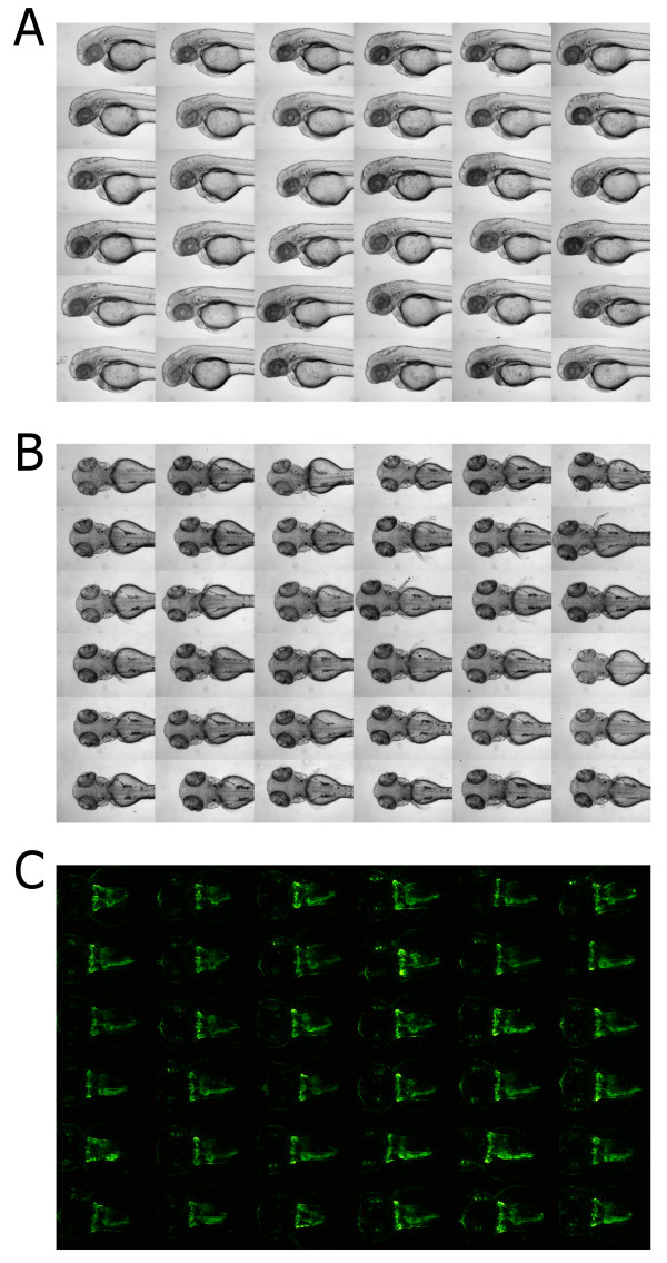
Figure 2.
Screening data obtained using the 3D printed orientation tools. Shown are illustrative examples of embryos within agarose cavities generated with 3D printed orientation tools. All images shown derive from single 96 well plates with laterally or dorsally oriented embryos, respectively (see also Additional file 3). (A, B) Cropped extended focus bright field images of 48 hpf zebrafish embryos: (A) lateral and (B) dorsal views. (C) Cropped maximum projections of deconvolved z-stacks of kidney regions of 48 hpf embryos of the Tg(wt1b:EGFP) transgenic line.

