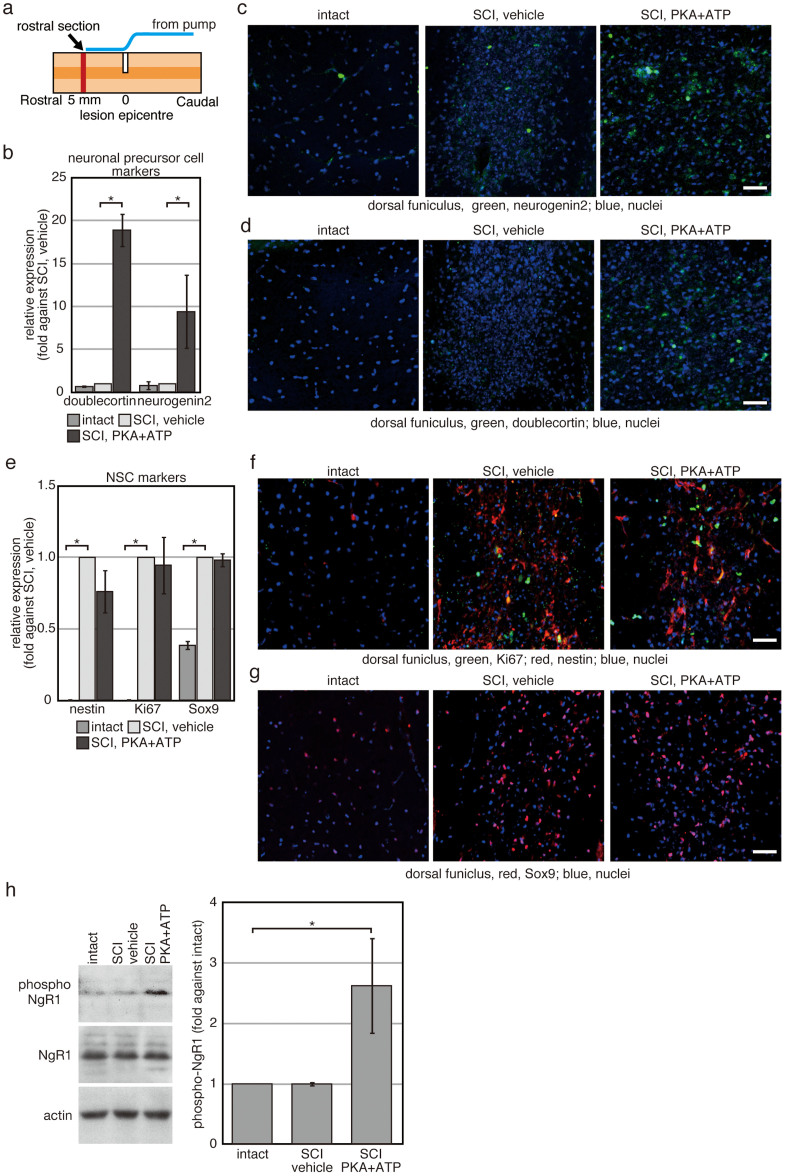
Figure 2. Extracellular treatment with PKA plus ATP increases the number of neuronal precursor cells in the peri-injury site.
Rats with SCI were treated with either vehicle or PKA plus ATP for 1 week. (a) Location of sections used in (c, d, f, g) is illustrated schematically. The sections were prepared from the spinal cord directly under the end-point of the tube from the infusion pump. (b–g) Transverse sections prepared from the spinal cord directly under the end-point of the tube from the infusion pump were used for immunofluorescence with indicated antibodies (c, d, f, g). Representative images of the dorsal funiculus were shown. The bars indicate 25 µm. In (b) and (e), signals from indicated antibodies were quantified by using ImageJ 1.48k (available at http://rsb.info.nih.gov/ij/). The average of the three independent animals was plotted and error bars indicate the S.D. (*p < 0.05, Student's t-test). (h) Western blotting analysis of proteins expressed in the spinal nerves. Cell extracts were prepared from the spinal cord at 1 week after SCI, and 40 µg of protein was analysed for each condition. Experiment was repeated for 3 times and intensities of signals were quantifyd by using Image J 1.48k. The average was plotted and error bars indicate the S.D. (*p < 0.05, Student's t-test).

