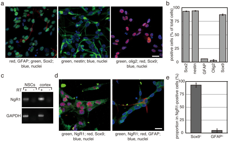
Figure 3. NSPs derived from the adult spinal cord express NgR1.
(a and d) After three rounds of neurosphere formation, NSPs were maintained as a monolayer culture. Cells were seeded onto plastic coverslips coated with polyornithine and laminin. After 2 days in culture, cells were fixed and used for immunofluorescence analysis with the indicated antibodies. The bars indicate 50 µm. (b) Cells expressing the indicated proteins were manually counted. DAPI-positive cells were also counted to obtain the total cell number. Each experiment was repeated four times, and more than 200 cells per sample were examined. Each bar and error bar represents the average cell number and the S.D., respectively. (c) Total RNA was prepared from cultured NSPs and used as the template for RT-PCR. Complementary DNA prepared from the adult rat cortex was used as a positive control. Glyceraldehyde 3-phosphate dehydrogenase (GAPDH) was employed as an internal standard. (e) Sox9-expressing cells and GFAP-positive cells were counted in NgR1-positive cells, respectively. They were indicated as percentage against the number of examined NgR1-positive cells. The results were obtained from 3 independent clones of NSPs and their average was shown. Experiments were repeated 3 times. Error bars indicate the S.D.

