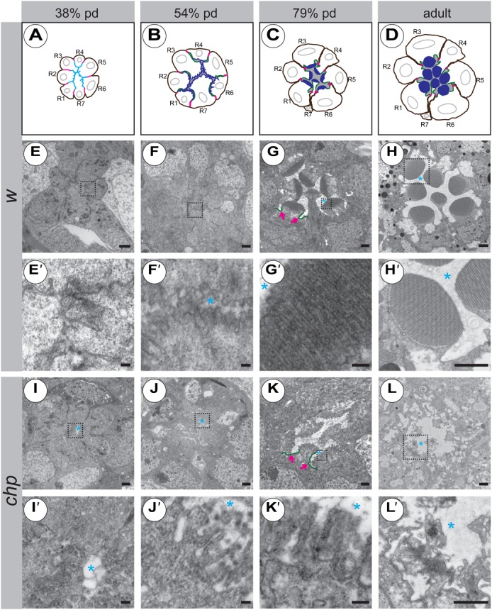
Fig. 4. Development of the apical compartment in chp mutant PRCs.

(A–D) Cartoons of different developmental stages in wild-type ommatidia (not taking into account the correct contacts made by individual rhabdomeres). Magenta: adherens junction. Cyan: common apical surface in panel A, as deduced by co-localisation of Crb and F-actin. Green: Crb, highlighting the stalk membrane. Blue: F-actin, highlighting the microvilli. Grey: interrhabdomerel space. R1–R7 = number of PRCs. pd = pupal development. (E–L′): Electron micrographs of tangential sections of wild-type (E–H′) and chp2 mutant (I–L′) ommatidia at 38% pd (E,E′,I,I′), 54% pd (F,F′,J,J′), 79% pd (G,G′,K,K′) and in the adult (H,H′,L,L′). The first defects in chp mutant ommatidia can already be detected at 38% pd, in that the microvilli are not as closely associated with each other as in wild type (compare panel E′ to panel I′). Scale bars: 1 µm (E–H,H′,I–L,L′) and 100 nm (E′–G′,I′–K′); blue asterisk, IRS; magenta arrows, stalk membrane (labelled in green).
