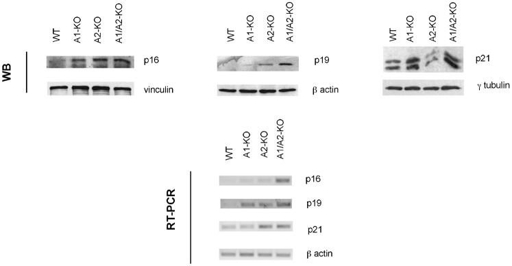
Fig. 6. Analysis of p16, p19 and p21 expression in WT, A1-KO, A2-KO and A1/A2-KO MEFs.

Expression of cell cycle inhibitors p16, p19 and p21 in representative MEFs from each genotype was determined by Western blot and RT-PCR at culture passage 7. β-actin gene expression was used as internal control. Vinculin, β-actin and γ-tubulin were used as loading control.
