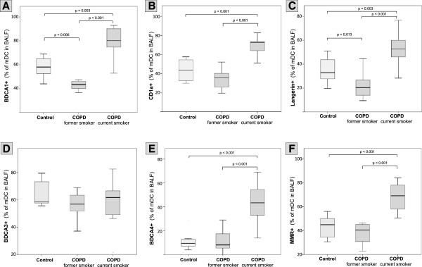
Figure 3.

Surface molecule expression on BALF mDCs. Boxplots show the expression (% positive mDCs in BALF) of BDCA-1 (A), CD1a (B), Langerin (C), BDCA-3 (D), BDCA-4 (E) and MMR (F) on mDCs in BALF from healthy never-smokers (Control), former smokers with COPD (COPD, former smoker) and current smokers with COPD (COPD, current smoker). Boxplots display the median (line within the box), interquartil range (edges of the box) and extremes (vertical lines). Outliers (all cases more distant than 1.5 interquartil ranges from the upper or lower quartil) were omitted in the graphs. Significant differences between two time groups are marked with the exact p-value.
