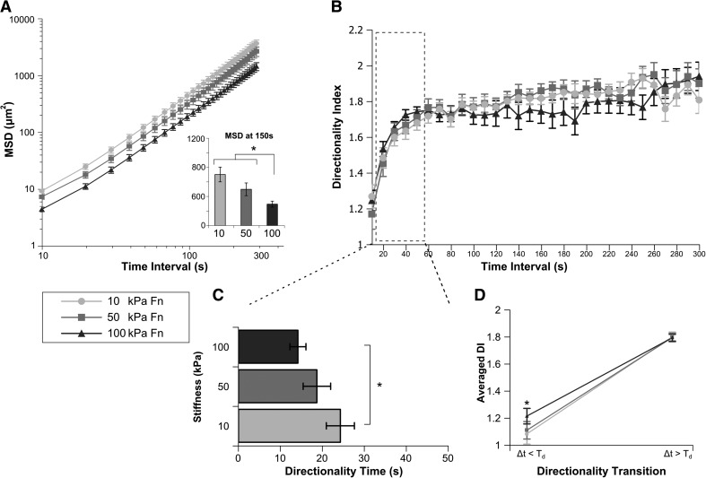
Figure 5. Neutrophils migrating on Fn-coated substrates toward fMLP show mechanosensitive differences in MSD, Td, and DI<.
Human primary glneutrophils migrating on Fn-coated gels of 10 kPa, 50 kPa, or 100 kPa stiffness toward a fMLP point source were tracked over a 30-min period (Supplemental Video 3). (A) The MSD of neutrophil migration paths is plotted as a function of time between steps. (Inset) The average MSD at a 150-s time interval for each condition. Cells migrating on 100 kPa Fn-coated gels show a significant decrease in MSD when compared with cells on 50 kPa or 10 kPa gels. *P < 0.05 100 kPa versus 50 kPa or 100 kPa. (B) The average DI of each condition is plotted over time interval. (C) For each cell, index DI(Δt) was fit to an exponential DI fit function, yielding mean values of Td. Cells migrating on Fn-coated gels show a stiffness-dependent change in Td, with cells migrating on 100 kPa gels transitioning at significantly shorter time scales to directed motion than those on 10 kPa gels. (D) The fit of DI(Δt) was also used to generate values for DI< and DI>. These data are plotted as the directionality transition. The DI< of cells migrating on 100 kPa gels is significantly more directed than cells on 10 kPa. DI> of cells on Fn-coated gels is independent of substrate stiffness. Error bars represent sem. *P < 0.05, 10 kPa versus 100 kPa.

