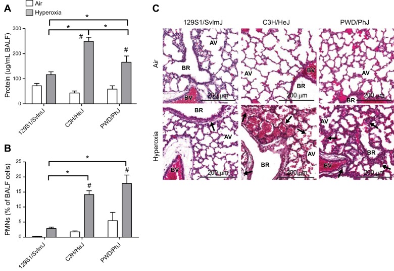
Figure 1.
Histopathologic and BALF responses to hyperoxia exposure in 129S1/SvImJ, C3H/HeJ, and PWD/PhJ neonates. A) BALF protein concentration. B) PMNs. C) Representative H&E-stained histopathology specimens from formalin-fixed neonatal lungs. Arrows show differences in peribronchiolar edema, vascular leakage, cell infiltrate, and alveolar septal thickness. AV, alveoli; BR, bronchus or bronchiole; BV, blood vessel. *P < 0.05 vs. indicated strain; #P < 0.05 vs. air-exposed control; Student-Newman-Keuls test.

