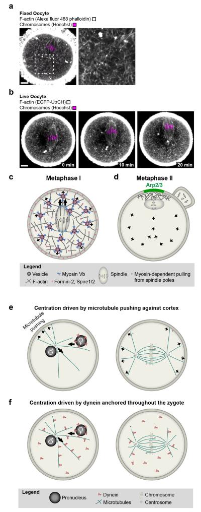
Figure 5. Transition from asymmetric to symmetric spindle positioning.
a | A cytoplasmic F-actin network mediates asymmetric spindle positioning during meiosis I in mouse oocytes. F-actin (grey; Alexa fluor 488 phalloidin) and chromosomes (magenta; Hoechst) in fixed mouse oocyte. Scale bar: 10 μm. b | Autofluorescence (green) around spindle region and chromosomes (magenta; Hoechst) in fixed mouse oocyte. Scale bar: 10 μm. c | F-actin (grey; EGFP-UtrCH) and chromosomes (magenta; Hoechst) in live oocyte during asymmetric spindle positioning. Scale bar: 10 μm. d | Model for vesicle-actin network mediated asymmetric spindle positioning during the first meiotic division e | Model for how the Arp2/3-complex helps to maintain the metaphase II spindle in cortical proximity while the egg awaits fertilization. f | Model for centration of pronuclei and first mitotic spindle by pushing of astral microtubules against the cortex. g | Model for centration of pronuclei and first mitotic spindle driven by dynein that is anchored throughout the cytoplasm.

