Fig. 6.
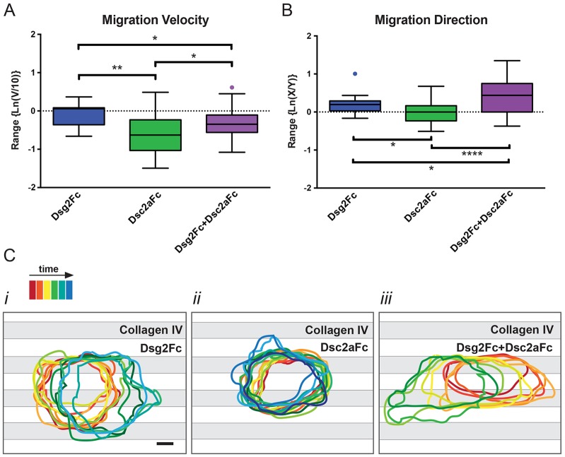
Desmosomal cadherin engagement regulates MDCK cell migration on dual-patterned substrates. The movement of single MDCK cells on each dual-patterned substrate condition was tracked by monitoring changes in nuclear position over time. (A) Cell migration rate (v) was normalized to single cell movement on only collagen IV surfaces, and plotted as a tukey box-and-whisker plot on a natural log scale [Ln(v/10 µm/h)]. The box marks the range from the 25th–75th percentile of the data and the band inside the box marks the median. Whiskers show the maximum and minimum values. (B) Directionality of movement was plotted as a tukey box-and -whisker plot on a natural log scale as a function of movement parallel to stripes (x) divided by perpendicular movement (y). *P<0.1; **P<0.01; ****P<0.0001. (C) Membrane activity during migration assays was assessed over time by tracing the cell contour from DIC images and projecting the outlines onto the stripe patterns (C). Statistical analysis used the Mann–Whitney, two-tailed t-test (Dsg2Fc, n = 13; Dsc2aFc, n = 31; Dsg2Fc+Dsc2aFc, n = 27).

