Fig. 3.
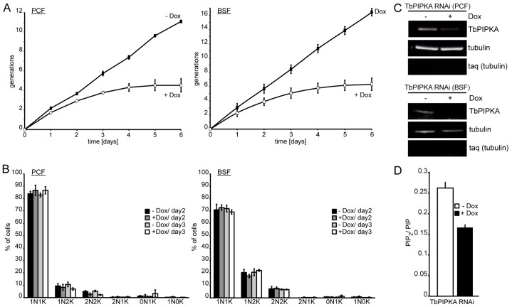
Depletion of TbPIPKA by RNAi. (A) TbPIPKA depletion (+Dox) caused a growth arrest in PCF and BSF cells. Data points are the means±s.e.m., n = 3. (B) Depletion of TbPIPKA does not cause a cell-cycle progression defect in PCF (left panel) or BSF (right panel) cells. Samples of control (−Dox) and induced-RNAi (+Dox) from PCF and BSF cells were taken after 2 and 3 days. Nuclear (N) and kinetoplast (K) DNA was labeled with DAPI to determine the cell cycle state. At least 300 cells were analyzed for each timepoint across three independent experiments. Values are mean±s.e.m., n = 3. 1N1K, one nucleus, one kinetoplast etc. (C) TbPIPKA mRNA depletion following induction of RNAi. RNA was isolated from PCF (top panel) and BSF (bottom panel) cells after growth in the absence or presence of 100 ng/ml doxycycline for 3 days. Tubulin served as the loading control, taq (tubulin) was used to determine DNA contamination. (D) Extracted lipids from TbPIPKA-depleted (+Dox) PCF cells were analyzed by HPLC and the PIP2∶PIP peak area ratio was plotted. Three independent experiments, values are mean±s.e.m.

