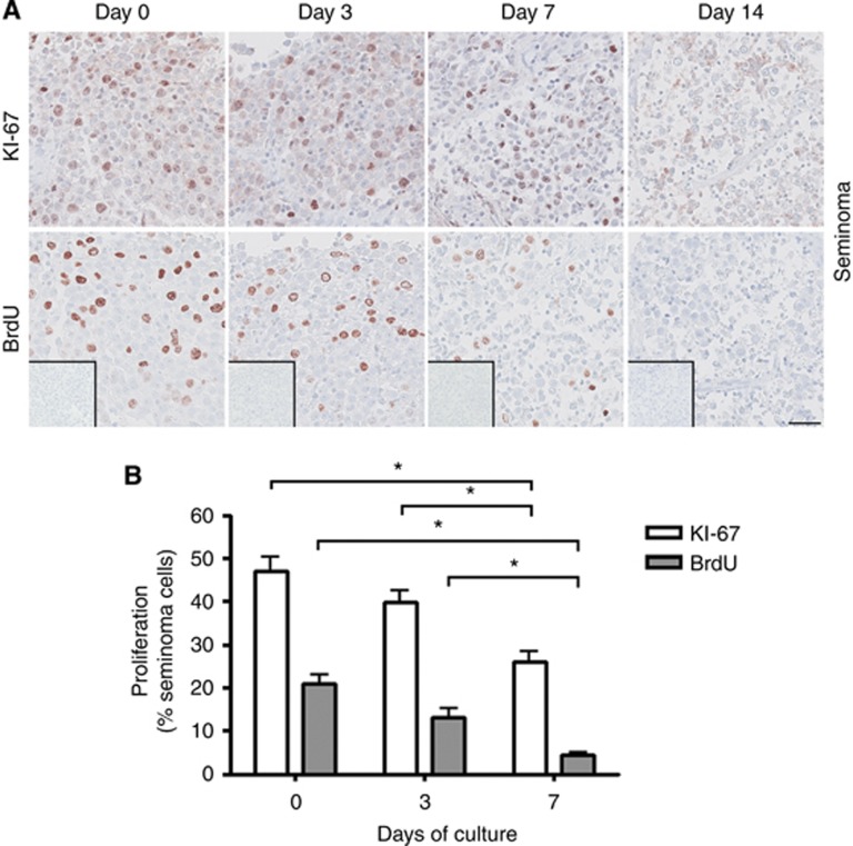
Figure 4.
Proliferation of seminoma cells in culture determined by KI-67 staining and BrdU incorporation assay after 0, 3 and 7 days of culture in the presence of 10% FBS. (A) Top panel shows IHC staining with KI-67. Bottom panel shows IHC staining with BrdU. BrdU staining detects incorporated BrdU during replication for 3-h period before tissue fixation. Inserts show negative control staining (no antibody). Serial sections of tissue counterstained with Mayer haematoxylin are shown in each column. Tissue from three patients was investigated, with 3 experimental replicates per time point per patient. Scale bar corresponds to 50 μm. (B) Quantification of proliferating seminoma cells (in percentage), determined by KI-67 staining and BrdU incorporation. Values represent mean±s.e.m. * indicates significant difference (P<0.05).

