Figure 2.
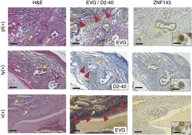
Representative pictures for H&E, elastica van Gieson (EVG), and immunohistochemical analyses of ZNF143and D2-40 in pleural involvement (pl) and lymphatic (ly) or vascular (v) invasion of moderately to poorly differentiated lung adenocarcinoma components (arrows; strong ZNF143+) (original magnification: × 100; inset: × 400). The EVG and D2-40 stainings very clearly reveal elastic fibres of the visceral pleura (pl(+)) or of the arterial medial wall (v(+)), and lymphatic endothelium (ly(+)), respectively (arrowheads). Bar=100 μm ( × 100) or 20 μm ( × 400).

