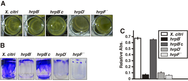
Figure 1.

Biofilm assays for X. citri, the hrp mutants and the hrpB−c strain. Representative photographs of biofilm formation assays for X. citri, hrp mutants and hrpB−c strains grown statically in 24-well PVC plates (A) or in borosilicate glass tubes (B) for seven days in XVM2 medium. (C) Quantification of biofilm formation by CV stain measured spectrophotometrically (Abs. at 600 nm). Relative Abs. indicates: CV Abs. 600 nm/Planktonic cells Abs. 600 nm. Values represent the mean from seven tubes for each strain. Error bars indicate the standard deviation.
