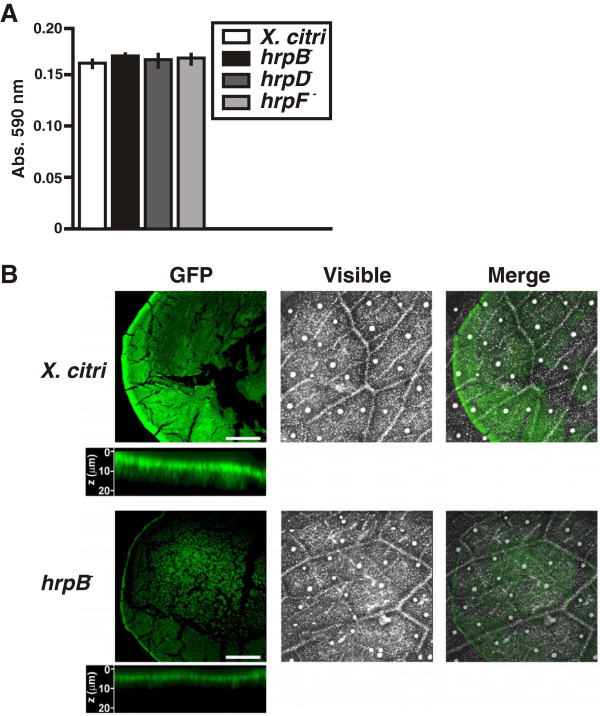
Figure 3.
Adherence of the hrp mutants to citrus leaf tissues and confocal laser scanning microscopy analysis on citrus leaves of X. citri and hrpB− strains. (A) Quantitative measurement of the CV retained by X. citri and hrp mutant strains adhered to abaxial leaf surfaces. Values represent the means of 20 quantified stained drops for each strain. Error bars indicate standard deviations. (B) Representative photographs of confocal laser scanning microscopy analysis of GFP-expressing X. citri and hrpB− cells grown on leaf surfaces. Below each of the fluorescent photographs of both strains, the ZX axis projected images accumulated over serial imaging taken at 0.5 μm distances (z-stack) are shown. Scale bars: 0.5 mm.

