Figure 8.

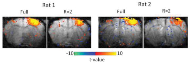
EPI-based fMRI maps of two rats responding to forepaw stimulation obtained from block-designed data with full and Rand+C6 (R=2) sampling patterns. Color maps were overlaid on the corresponding baseline EPI images.

EPI-based fMRI maps of two rats responding to forepaw stimulation obtained from block-designed data with full and Rand+C6 (R=2) sampling patterns. Color maps were overlaid on the corresponding baseline EPI images.