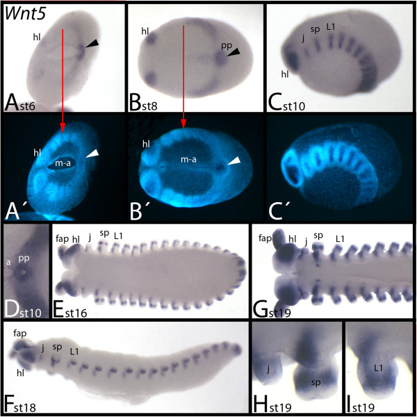
Figure 6.
Expression of E. kanangrensis Wnt5. Anterior is to the left. (A) Ventral view. Red arrow marks the anterior border of expression (of the broad domain; note additional expression in the head lobes). Arrowhead indicates strong expression in the posterior pit region. (B) Ventral view. Arrow and arrowhead as in (A). (C) Lateral view. (D) Ventral view. Close-up of the posterior of the embryo of (C). Note the strong expression in a ring around the centre of the posterior pit region. (E) Ventral view. (G) Ventral view. Anterior part of an embryo. (F) Lateral view. (H) Close-up of a jaw and a slime papilla. (I) Close-up of a walking limb. (A′), (B′) and (C′) represent DAPI staining of the embryos of (A), (B) and (C). a, anus; fap, frontal appendage; hl, head lobe; j, jaw; L1, first walking-limb-bearing segment; m-a, mouth-anus furrow; pp, posterior pit region; sp, slime papilla; st, stage.

