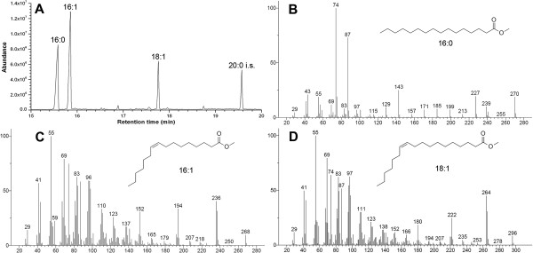
Figure 3.

Identification of free fatty-acid compositions of the AtFatA-expressing strain by gas chromatography-mass spectrometry (GC-MS). (A) A total ion chromatogram (TIC) of free fatty-acid methyl esters from E. coli BL21 star(DE3) harboring pA-AtFatA after being induced for 16 h. (B) Mass spectrum of palmitate methyl ester (16:0); (C) mass spectrum of palmitoleate methyl ester (16:1Δ9); (D) mass spectrum of cis-vaccenate methyl ester (18:1Δ11). The structures were matched by searching a standard NIST library. The molecular ion peaks of these fatty-acid methyl esters are marked with circles.
