
Figure 4.
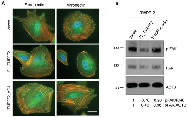
TMEFF2 reduces stress fiber and focal adhesion formation and activation. A) RWPE2 cells expressing FL_TMEFF2, TMEFF2_ΔGA, or the vector were cultured on cover glass coated with fibronectin or vitronectin for 3 h and then stained with anti-vinculin (green), rhodamine phalloidin (orange), and DAPI (blue). Scale bar, 20 μm. B) Immunoblotting of phosphorylated FAK (Y397), FAK and β-actin (ACTB) in RWPE2 cells. Numbers under the western blots represent the densitometry quantifications using ImageJ.