Figure 4.

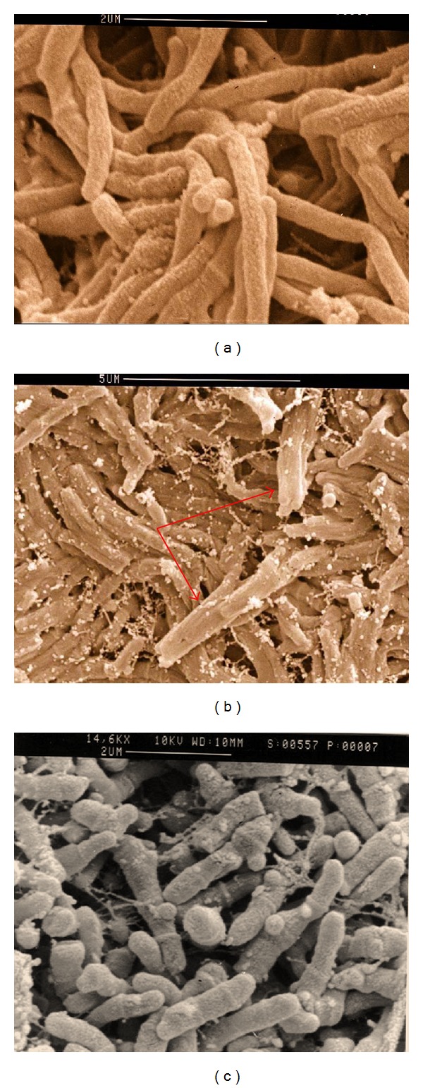
Scanning electron microscopy of M. farcinogenes (a), M. senegalense (b), and Nocardia farcinica (c). Note the true-nonfragmenting branched filaments in both species and the presence of “synnemata” in M. senegalense (arrow).

Scanning electron microscopy of M. farcinogenes (a), M. senegalense (b), and Nocardia farcinica (c). Note the true-nonfragmenting branched filaments in both species and the presence of “synnemata” in M. senegalense (arrow).