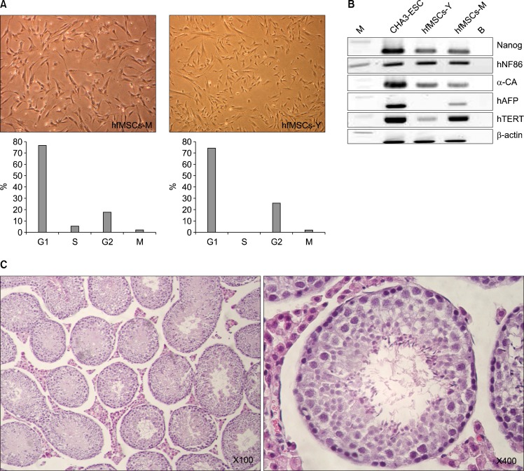
Fig. 1.
Morphology of hfMSCs derived from fetal membranes (hfMSCs-M) and the yolk sac (hfMSCs-Y). hfMSCs were assessed at passage number 8 to 10. (A) The morphologies of hfMSCs including hfMSCs-M and hfMSCs-Y were similar to the round-spindle shape of mesenchymal stem cells (Upper, ×100). Cell cycle analysis of hfMSCs showed increased S/G2 phase, indicating proliferative activity (Lower). (B) RT-PCR analysis for stem cells markers in embryonic stem cells (CHA3-ESC), hfMSCs-Y and hfMSCs-M. (C) Histological analysis for teratoma formation in the testicular capsules of 8-week-old NOD/SCID mice after hfMSCs transplantation.

