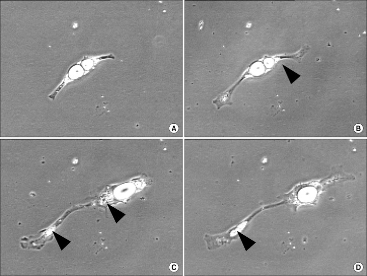
Fig. 1.
Pig-derived mature adipocyte proliferation in vitro. Basic differences were observed with beef-derived mature adipocytes. The culture method for both the beef and the pig cell systems were as described previously by Fernyhough et al. (3–5). Pig perirenal adipose tissue was enzymatically dispersed, and the lipid-laden fat cells were subjected to ceiling culture and eventually purified using two serial differential plating within 4 d of initial isolation. The purified mature adipocytes were exposed to DMEM/F12+10% FBS medium and monitored daily for morphological changes in the fat cells. Panel (A): the appearance of a bi-locular adipocyte in ceiling culture at 48 h after initating the second ceiling culture. The cell extended and attached firmly to the cultureware. Panel (B), (C), (D): At day 3, 5, 7 (respectively) after initiating the second ceiling culture, the cytoplasm stretched as the cell elongated. A small portion of cellular lipid was observed moving from the central part of the cell to one end (black arrowheads). Panel (E), (F), (G): At day 9, 10, and 11 (respectively) most of the cellular lipid was extruded out of the cell (white arrows). Panel (H): On day 12 after ceiling culture, the cell divided into two daughter cells (asterisks) after expelling a large amount of cellular lipid. All photomicrographs were captured at 20X magnification with a NIKON inverted Diaphot microscope equipped with Sony RGB (0.6 in chip) camera and OPTIX image analysis system.


