
Figure 2.
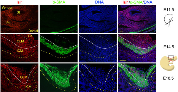
Double immunostaining for Isl1 and α-smooth muscle actin in mouse smooth muscle cells of the dorsal pylorus. Isl1 and α-SMA co-expression in smooth muscle cells at E11.5, E14.5, and E18.5. Yellow dotted lines mark the epithelial basement membrane, white thick dotted lines indicate ICM and OLM boundary, and white dotted lines indicate OLM and pancreas boundary. Red staining is Isl1, green staining is α-SMA, and DAPI nuclear counterstaining (DNA) is blue. α-SMA, α-smooth muscle actin; ICM, inner circular muscle; OLM, outer longitudinal muscle; Pa, pancreas; St, stomach. Scale bars: 50 μm.