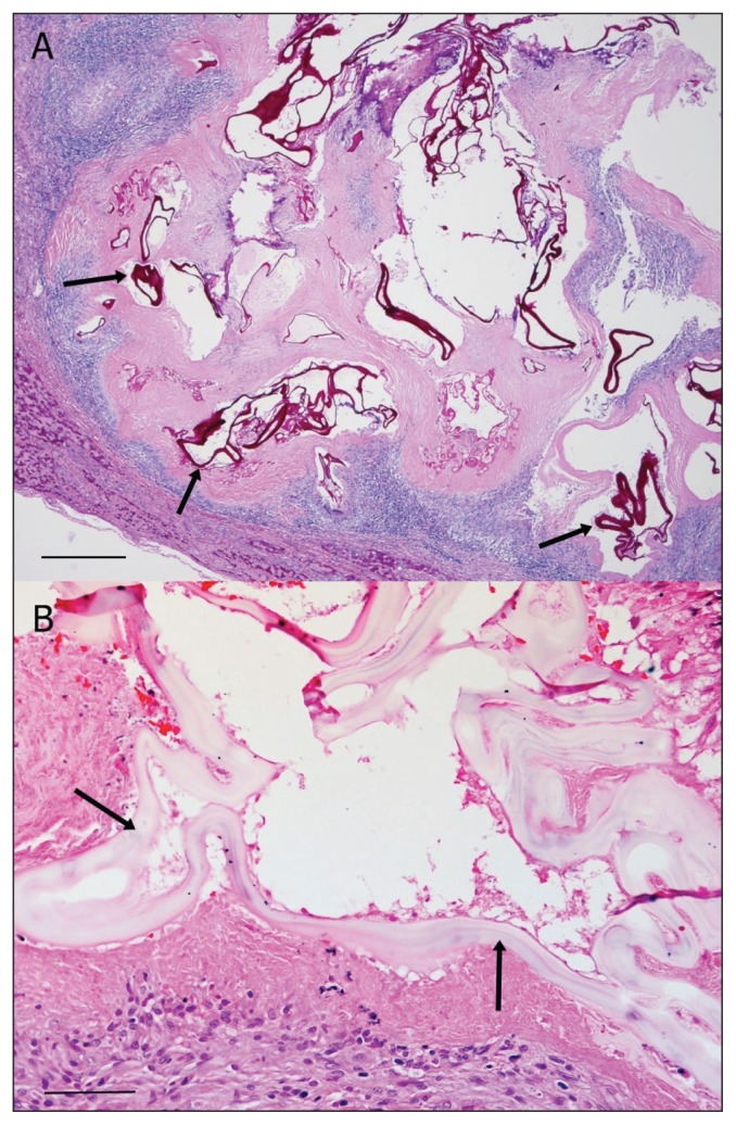
Figure 2.
Photomicrographs of a section of cystic tissue from the mass shown in Figure 1B. A — Note degenerating cysts in the liver containing Periodic acid-Schiff (PAS)-positive hyaline membranes (arrows) surrounded by eosinophilic granulomatous inflammation and necrosis. Stain = PAS, Bar = 250 μm. B — Note degenerating cysts in the liver that are lined by a hyaline membrane (“laminated layer”; arrows). Stain = hematoxylin and eosin, Bar = 50 μm.

