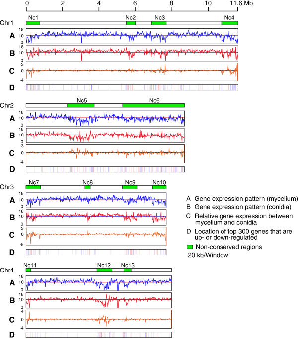
Figure 5.

Comparison of gene expression patterns between mycelium and conidia on the chromosomes of Fusarium graminearum . (A) Each chromosome was divided into 20 kb windows. Log2-transformed read coverage per window was used to quantify gene expression levels in mycelium of F. graminearum. (B) Similarly, the Log2-transformed read coverage per window was used to quantify gene expression levels in conidia of F. graminearum. (C) The ratio of the log2-transformed RPKM values per window between mycelium and conidia. (D) The locations of the 300 genes that show the strongest up-regulation comparing mycelium to conidia (red lines) and the 300 genes that show the strongest down-regulated (blue lines) on each chromosome of F. graminearum. Green boxes represent non-conserved regions in F. graminearum.
