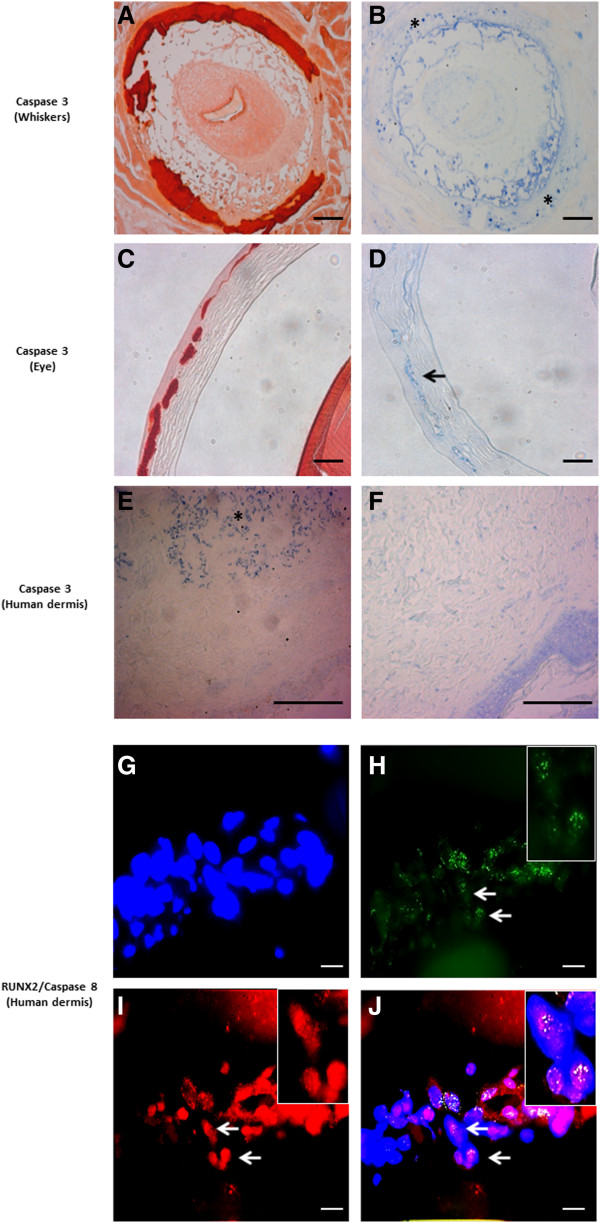
Figure 9.
Immunohistochemical staining results for Caspase 3 and Caspase 8. Upper panel: Staining is performed on whisker and eye sections from the Abcc6 knockout mouse (A-D, ×10) and human skin (E, F, ×20). Alizarin Red stains was used to localize mineralization (A, C). Positive staining for Caspase 3 is obtained on murine whiskers and eye (B, asterisk; D, arrow), co-localizing with ectopic mineralization. In human PXE dermis, Caspase 3 stains positive in the middermis (E, asterisk), while no staining is noted in controls (F). Scale bar = 100 μm. Lower panel: Fluorescent immunohistochemistry for Caspase 8 and RUNX2 confirms co-localisation in PXE skin (J, white arrow). DAPI nuclear staining (G). Merged RUNX2-DAPI image ((H, green fluorescence indicates RUNX2 staining, arrow) and merged Caspase 8-DAPI image (I, red fluorescence indicates Caspase-8 staining, arrow). (n = 5 each). Scale bar = 20 μm.

