
Figure 7.
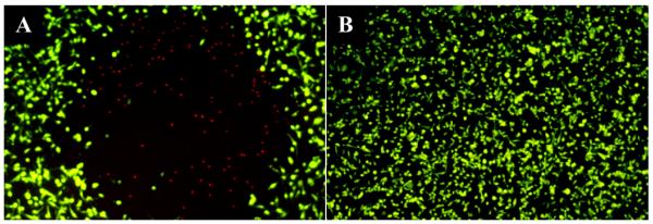
Fluorescence images of BT549 cells incubated with: (A) S30-DTTC@SiO2-MB and (B) S30-DTTC@SiO2 after 1 hr of laser irradiation, taken with a 5× objective. Cell death is only seen after treatment using MB-encapsulated particles, and is limited to the laser irradiated area. The green color is from hydrolyzed FDA, indicating live cells. The red color is from DNA intercalated PI that only enters cells with compromised membranes, indicating dead cells.