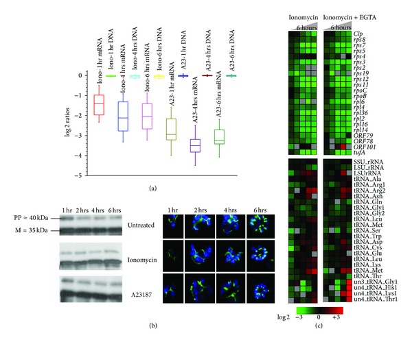
Figure 2.

Apicoplast development and DNA replication are hardly inhibited by calcium ionophores. (a) Apicoplast DNA replication is not inhibited by calcium ionophores. Box plots for the log2 expression ratios of averaged oligos representing all apicoplast genes (obtained from the microarray hybridization results filtered for 3-fold change in 2 time points) and log2 ratios of the treated and untreated apicoplast DNA (obtained from a CGH experiment where the total DNA from the treated parasites was hybridized against the total DNA from the untreated parasites) have been compared. (b) Western blots show the processed band (≈35 kDa) of the ACP-GFP fusion protein (≈40 kDa) directed towards apicoplast indicating that the protein has been imported and processed without much interference even after 6 hours after treatment. Fluorescence microscopy done on ACP-GFP expressing parasites confirms the western results. (c) Relative expression of apicoplast genes in ionomycin treatment of schizonts both in presence and absence of 3 mM EGTA. (I-ionomycin, I + E-ionomycin plus EGTA).
