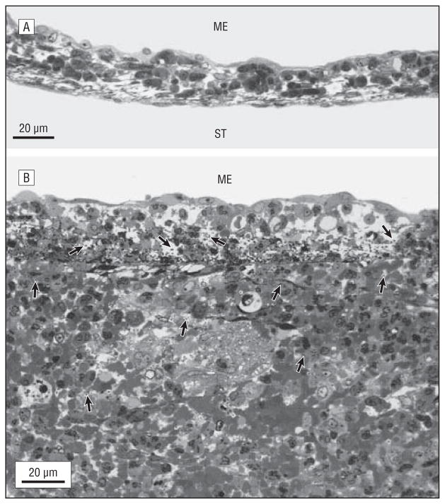
Figure 1.
Inflammation of the round window membrane (RWM) (toluidine blue, original magnification ×600). A, The most severe pathological changes observed in an RWM from the apolactoferrin-treated group, showing a slightly elevated thickness and a small number of inflammatory cells. B, The most severe pathological changes seen in the RWM of the phosphate-buffered saline–treated group, showing a large number of bacteria and inflammatory cells in the RWM and a substantial increasing of its thickness. Bacteria (arrows) are seen within the RWM and adjacent scala tympani (ST). ME indicates middle ear; arrows indicate bacteria.

