
Figure 2.
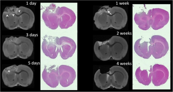
Evolution of controlled cortical impact injury over time.Ex vivo magnetic resonance imaging (MRI) and corresponding hematoxylin and eosin stains demonstrate the controlled cortical impact (CCI) lesion’s extension from the cortex through the corpus callosum. By 4 weeks postinjury, the lesion had evolved into a cavity, with minimal further changes observed by 8 weeks (data not shown). Arrowheads on day 1 MRI scans indicate areas of edema. The arrow on the day 7 scan indicates an area of hemorrhage.