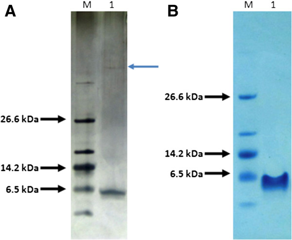
Figure 4.
Purified p53pAnt peptide. SDS-PAGE 10% T, 3% C; Tris-Tricine buffer system. M: Molecular weight marker; 1: p53pAnt. A: 0.125 μg of peptide visualized with silver staining. Blue arrow indicates a contaminant protein band. B: 2 μg of peptide visualized with Coomasie staining. The contaminant protein band is not visualized.

