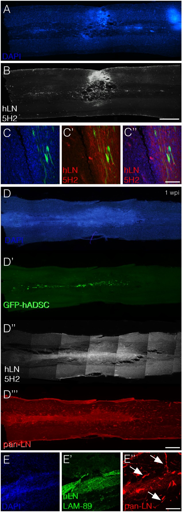
Figure 6. hADSCs secrete laminin in the spinal cord independently of SCI.

A–C) Horizontal section of an animal subjected to spinal compression and injected with GFP-hADSCs one week after injury/injection. Panels A and B show photomontages depicting DAPI staining (A) and immunoreactivity for anti-human laminin (B). Note that cell infiltrates and laminin are located in corresponding regions. Panel C shows confocal images demonstrating the coincident localization of GFP-positive hADSCs (green), cell infiltrates (blue) and human laminin (red). D–E) Horizontal section of an uninjured animal one week after transplantation with GFP-hADSCs. Panels D to D’’’ show photomontages depicting DAPI staining (blue, D), GFP-positive hADSCs (D’, green), immunoreactivity for anti-human laminin (D’’, white) and immunoreactivity for anti-pan laminin (D’’’, red). Panel E shows confocal images to demonstrate that while the anti-pan laminin antibody labels rat blood vessels (E’’, red), the anti-human laminin does not (E’). Bars: A, B, D = 1 mm, C = 100 µm, E = 200 µm.
