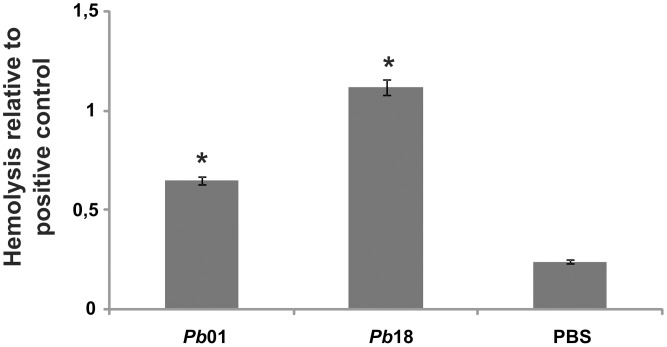
Figure 4. Hemolysis of sheep erythrocytes in the presence of Paracoccidioides yeast cells.
Pb01 and Pb18 107 yeast cell suspensions were incubated with 108 sheep erythrocytes for 2 h at 36°C in 5% CO2. As a negative or positive control, respectively, erythrocytes were incubated with phosphate buffered saline solution (PBS) or sterile water. The optical densities of the supernatants were determined with an ELISA plate reader at 405 nm. The experiment was performed in triplicate, and the average optical density of each condition was used to calculate the relative hemolysis of the experimental conditions or the negative control against the positive control. The data are plotted as the mean ± standard deviation. *: statistically significant difference in comparison with PBS values according to Student's t-test.

