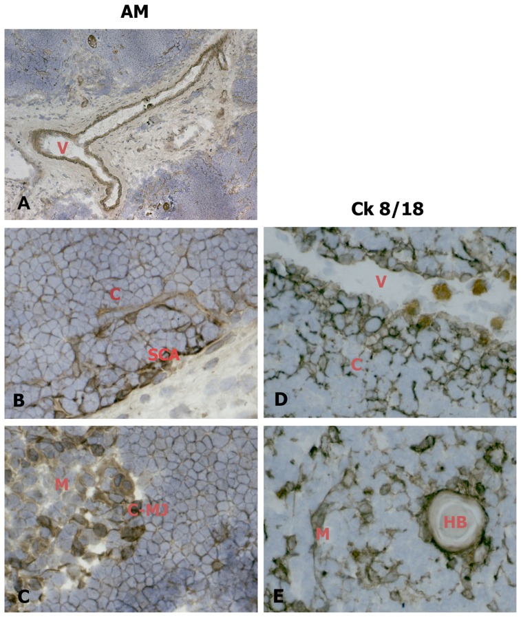
Figure 1. AM distribution in the epithelial compartment in cryostat sections of human newborn thymic tissue.
Sections were incubated with an antibody against AM (left column) or Ck 8/18 (right column). AM expression in blood vessels (A). AM distribution is similar to that of Ck8/18 positive epithelial cells in the cortex (B, D), and the medulla (C, E). When, for control purposes, the primary antibody was replaced by a non-immune normal goat serum or by primary antibodies pre-absorbed with antigen excess, no reactivity was observed (not shown in the figure). Original magnification ×50 A; ×400 B, C, E, F. V: blood vessel; C: cortex; M: medulla; SCA: subcapsular area; C-MJ: cortico-medullary junction; HB: Hassal's body.

