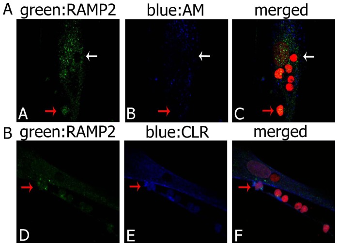
Figure 8. RAMP2, CLR and AM distribution in cultured thymocytes.
(A) Immunofluorescence staining of thymocytes co-cultured with TECs for RAMP2 (green) and AM (blue). Only some thymocytes express RAMP2 and AM, mainly in the cytoplasmic compartment (one of them indicated by a red arrow), whereas other cells are not immunostained (white arrow). (B) Immunofluorescence staining of thymocytes co-cultured with TECs for RAMP2 (green) and CLR (blue). Co-presence of green and blue fluorescence is evident only in some thymocytes (red arrow). Cell nuclei are red stained with propidium iodide. When, for control purposes, the primary antibody was replaced by a non-immune PBS solution, no reactivity could be observed (not shown in the figure).

