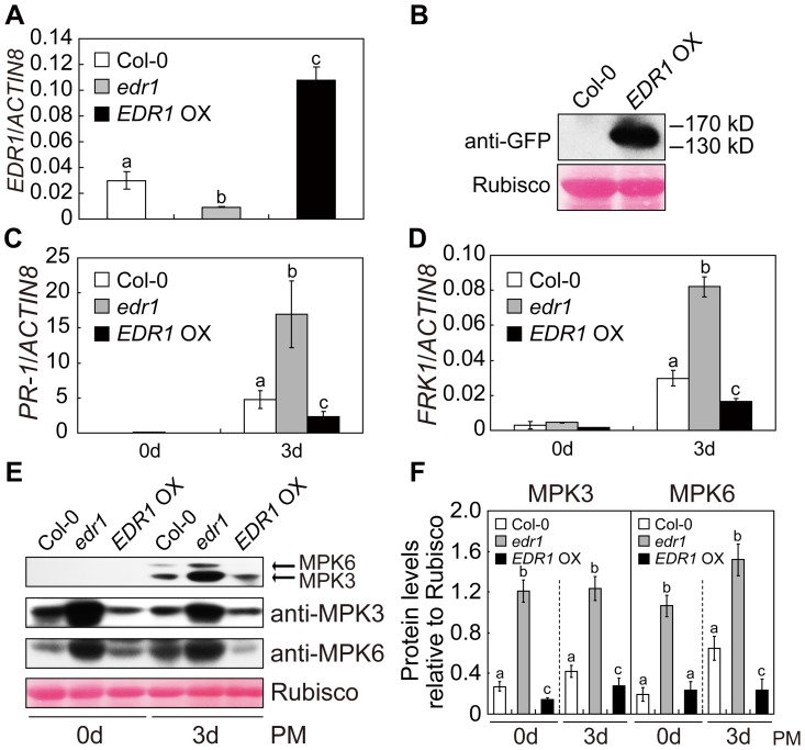
Figure 2. Over-expression of EDR1 reduced the kinase activity and protein levels of MPK3 and MPK6.
(A) The transcript accumulation of EDR1 was examined by quantitative real-time RT-PCR for wild-type Col-0, edr1 and EDR1 over-expressing plants. ACTIN8 was used as an internal control. Error bars represent the standard deviation of three biological replicates. Different letters represent statistically significant differences (P<0.05, one-way ANOVA). (B) EDR1-GFP is properly expressed in EDR1 over-expressing plants. Immunoblotting was performed using anti-GFP antibody in four-week-old plants. The large subunit of Rubisco is shown as a protein loading control. (C–D) The transcript accumulation of PR-1 (C) or FRK1 (D) was examined by quantitative real-time RT-PCR. Leaves from Col-0, edr1 and EDR1 over-expressing plants after infection by powdery mildew for 0 d and 3 d were collected for RNA isolation. Error bars represent the standard deviation of three biological replicates. Different letters represent statistically significant differences (P<0.05, one-way ANOVA). (E) Col-0, edr1 and EDR1 over-expressing plants were infected by G. cichoracearum. Immunoblot was performed using anti-pTEpY, anti-MPK3 and anti-MPK6 antibodies, as indicated. The large subunit of Rubisco is shown as a protein loading control. The experiment was repeated three times with similar results. PM: powdery mildew infection. (F) The protein bands of MPK3 and MPK6, as well as Rubisco, were quantified with ImageJ. The protein levels of MPK3 and MPK6 in each sample were evaluated by comparing to Rubisco. The error bars represent the standard deviation of three biological replicates. Different letters represent statistically significant differences (P<0.05, one-way ANOVA). PM: powdery mildew infection.

