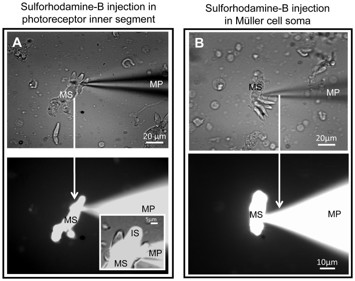
Figure 5. Unidirectional flow of sulforhodamine-B dye from cone to caiman Müller cell.
(A) Upper panel: whole-cell patch clamp technique using micropipette (MP) filled with sulforhodamine-B penetrating the inner segment of a cone attached to a Müller cell. The dye filled the cone and the Müller cell: tracing of the photoreceptor cell body and inner segment (IS) and spreading throughout the whole Müller cell from the soma (MS) to the endfeet. Lower panel: combined (DIC and fluorescent) image showing that sulforhodamine-B is not permeable to neighboring photoreceptors, but filled only the cone which was patched. Insert shows an enlarged image of fine attachments of three cones to a single glial cell where two cones are not showing fluorescent dye. (B) Upper panel: whole-cell patch clamp of a Müller cell resulted in the dye-tracing of the cell body and endfeet, while no spreading of the dye occurred to the photoreceptors attached to this Müller cell. Lower panel: patch of the Müller cell soma only traced the Müller cell; the dye did not spread to the attached photoreceptors.

