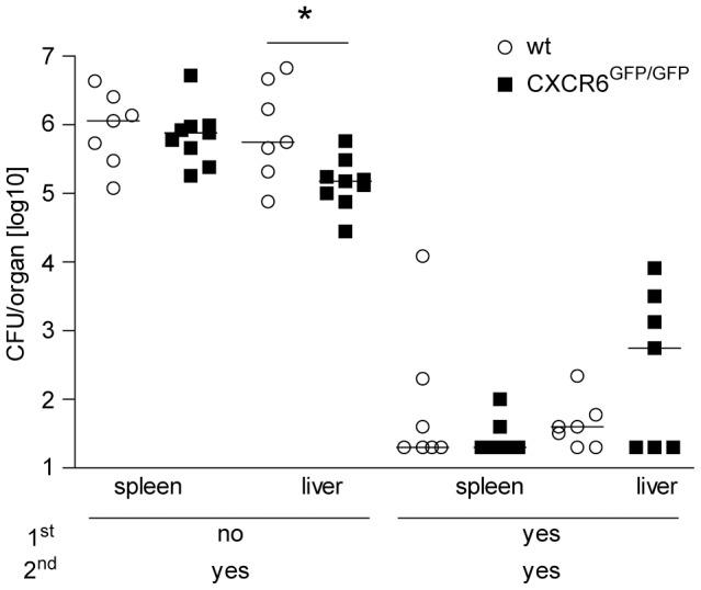
Figure 6. Control of secondary L. monocytogenes infection in CXCR6-deficient mice.

Wt and CXCR6GFP/GFP mice were primary infected with 5×10³ Lm i.v. and 60 days later challenged with 1×10⁵ Lm i.v. Two days after secondary infection, listeria titers in spleen and liver were determined. Colony forming units (CFU) for individual mice and the median of one representative experiment of two are shown, n≥7. Detection limit was 20 CFU. *, p<0.05.
