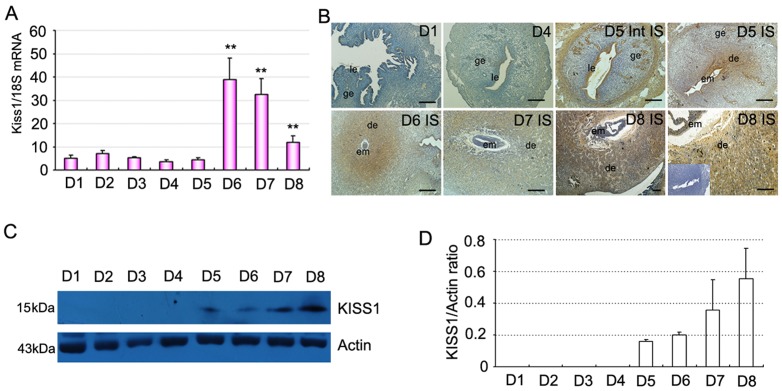
Figure 1. Expression of Kiss1 in mouse uterus during early pregnancy.
(A) Levels of Kiss1 mRNA expression in the uterus during early pregnancy via quantitative PCR analysis. (B) Immunostaining of KISS1 protein in mouse uterus on days 1, 4, 5, 6, 7, and 8 of pregnancy. Inset: control (without primary antibody). (C) Representative immunoblotting results for KISS1 protein in mouse uterus on days 1–8 (D1–D8) of pregnancy. (D) Densitometric analyses of KISS1 protein in mouse uterus on days 1–8 of pregnancy. All experiments were repeated three times. Data are shown as means ± SEM. **p<0.01 compared with D1 of pregnancy. Scale bar, 50 µm. IS, implantation site; Int IS, inter-implantation site; em, embryo; ge, glandular epithelium; le, luminal epithelium; de, deciduas.

