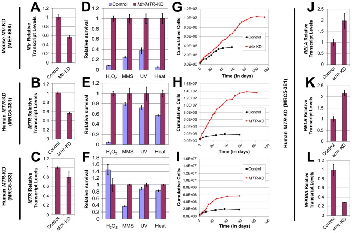
Figure 4. Effects of genetic Meth-R on stress tolerance, replicative lifespan and the expression of NFκB signaling genes in cultured mammalian cells.
(A–C) qRT-PCR determination of the extent of methionine synthase (Mtr/MTR) transcript depletion in cultured (A) mouse embryonic fibroblasts (MEF-688), as well as (B) human MRC5-381 and (C) human MRC5-383 fibroblasts (C). Transcript levels are normalized to those in mock-infected control cells. (D–F) Relative post-stress survival of methionine synthase knockdown fibroblasts (purple) (D) MEF-688, (E) MRC5-381, and (F) MRC5-383, as compared with mock-infected controls (light blue) (values shown are computed relative to non-stressed cell viability, and normalized to survival of Mtr/MTR-KD strains). (G–I) Replicative lifespan analyses of methionine synthase knockdown strains (G) MEF-688, (H) MRC5-381, and (I) MRC5-383, as compared with mock-infected controls. (J–L) qRT-PCR determination of (J) RELA, (K) RELB and (L) NFKBIA transcript levels in MTR-knockdown human fibroblasts (MRC5-381), as compared with transcript levels in mock-infected control cells. For panels A–F and J–L, bars denote SEM.

