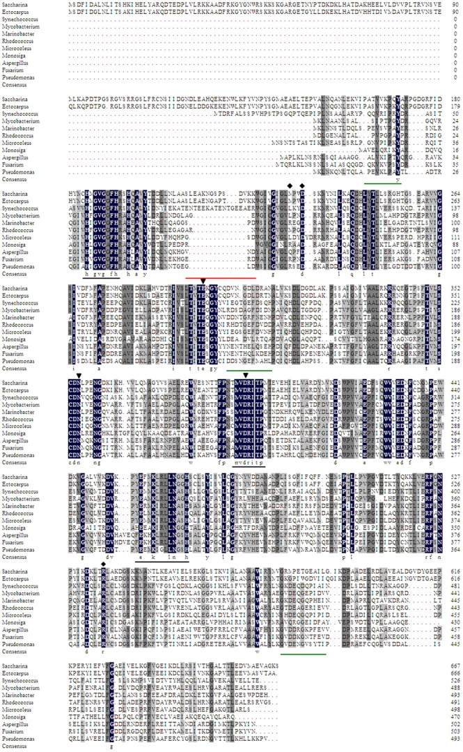
Figure 2. Multiple sequence alignment of MDHs from typical species of Phaeophyceae, Cyanobacteria, Actinobacteria, Proteobacteria, Ascomycota and Codonosigidae.
Identical residues among all MDHs were shown in black boxes. The representative conserved regions among PSLDRs were underlined in black. The deletions of β-sheets in SjM2DH were underlined in green while the extra anti-parallel β-sheet was underlined in red. ▾, residues for substrate-binding; ⧫, residues for NADH-binding. The accession numbers were listed in File S2.

